A Bone Marrow Biopsy Names the Cell, Not Your Future: How to Read the Report
A bone marrow biopsy takes two samples through one needle site and produces one of the strangest documents in medicine. Part of it comes back in days. Part takes weeks. Each part reads like its own verdict. A cellularity percentage, a blast count, a fibrosis grade, a flow cytometry panel, a karyotype, a list of mutated genes. None of those lines is the diagnosis. The marrow report is where several separate tests get bound into one answer, and the answer only holds if someone reads them together, the discipline Healz was built on.
This post walks the report from the top: why marrow gets sampled, what the aspirate sees that the core biopsy cannot, what each number measures, and what turns a frightening word into a diagnosis. A scary term in a findings section is a finding. The conclusion sits at the bottom, and it is written last for a reason.
Why anyone takes marrow in the first place
Blood tells you what the factory shipped. Marrow tells you what the factory is doing. When counts are low or strange and the blood cannot explain itself, the explanation lives upstream. The usual reasons to sample are unexplained cytopenias, suspected leukemia, lymphoma, myelodysplastic syndrome, or a plasma cell disorder (per StatPearls). It is also used to stage a known cancer and to check response after treatment (per ICSH). In adults the sample comes from the posterior iliac crest, the back of the hip bone, under local anesthetic that numbs skin and bone surface.
Not every suspicion needs it. For Hodgkin lymphoma the Lugano classification changed practice: if an FDG PET/CT is done, a marrow biopsy is no longer indicated for routine staging, and in diffuse large B-cell lymphoma it is needed mainly when the PET is negative and discordant histology would change management (Lugano, 2014). When a team does send you, the question is about the marrow's own cells. Myeloma is the clear example: diagnosis requires 10% or more clonal plasma cells in the marrow or a biopsy-proven plasmacytoma, and 60% or more is by itself a myeloma-defining event (IMWG criteria). Our piece on the vague symptoms that hide multiple myeloma covers how people arrive there.
Ask Healz.
One chat instead of ten apps. 1M+ rare cases checked. The root cause, not the label. Private.
The two samples, and why one alone is not enough
The aspirate is the liquid draw. It gives cell detail: what individual cells look like, how they are maturing, and the differential count, performed on 500 nucleated marrow cells alongside 200 white cells on the blood smear (WHO methodology). It is also what the downstream tests need, because flow cytometry, chromosome culture, and molecular panels all want living cells in suspension.
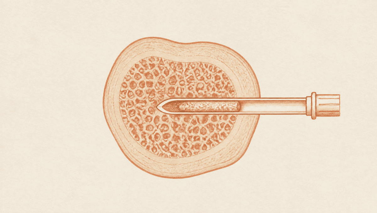
The core biopsy, also called the trephine, is a solid cylinder of bone and marrow, roughly 2 mm wide, and ICSH says the core from an adult should be at least 2 cm long, with guidance putting the workable minimum at 1.5 to 2 cm so the section holds at least 10 partially preserved intertrabecular areas (ICSH 2008). It answers what the liquid cannot: overall cellularity, assessed better on the core, plus fibrosis, architecture, and the patchy infiltrates that cling to the trabeculae and never make it into a syringe, lymphoma among them. And when the aspirate is a dry tap, meaning nothing could be drawn, the core becomes mandatory, because a dry tap usually means the marrow is fibrotic, packed, or infiltrated rather than that the operator missed.
One honest limit belongs here. A biopsy samples one small site, and in lymphoma involvement is often focal: in bilateral-biopsy series, 12% to 35% of patients who had marrow involvement were positive on only one side, though because marrow involvement is uncommon overall, the second biopsy changed the stage in only about 1% to 6% of all patients staged. At least one large modern series found no benefit from bilateral sampling. A negative marrow is evidence, not proof.
What the numbers on the page are actually measuring
Cellularity is the share of marrow space occupied by blood-forming cells rather than fat. The old bedside rule is 100 minus your age, and it is rough: a large study of United States adults put the 90% reference interval at 30% to 75% across ages 18 to 90, declining roughly 3% per decade, so the classic rule reads a normal older marrow as hypocellular (American Journal of Clinical Pathology, 2024). Packed above expectation suggests proliferation. Far below raises failure, and under 25% is part of how severe aplastic anemia is defined.
Myeloid to erythroid ratio compares white to red cell precursors, normally 2:1 to 4:1 (per StatPearls, Laboratory Evaluation of Bone Marrow), with published ranges varying between sources, and a shift says which line is running hot or has stalled.
Blast percentage is the line people search for first. Blasts are the youngest precursor cells, normally under 5% of marrow nucleated cells in adults, and at 20% or more the diagnosis is acute leukemia. The WHO 5th edition removed that fixed requirement for acute myeloid leukemias with a defining genetic abnormality, BCR::ABL1 and mutated CEBPA excepted, while the 2022 International Consensus Classification sets a 10% floor. Two systems, one point: the genetics can outrank the count.
Dysplasia means the cells look malformed, and it only counts above a threshold. At least 10% of cells in a lineage must be dysplastic to support a myelodysplastic diagnosis, a cutoff chosen to exclude the mildly odd-looking cells found in healthy older people (WHO). Reactive causes have to be excluded first.
Ring sideroblasts, red cell precursors with iron circling the nucleus, show up on a Prussian blue iron stain. Under the WHO revised 4th edition, myelodysplastic syndrome with ring sideroblasts required 15% or more, or 5% or more when an SF3B1 mutation was present. The WHO 5th edition inverts that logic: the entity is defined by the SF3B1 mutation, and 15% or more ring sideroblasts substitutes when SF3B1 is wild type or unknown.
Fibrosis grade runs MF-0 to MF-3 on the European consensus scale (Thiele et al., Haematologica 2005), from scattered reticulin with no intersections to dense reticulin with coarse collagen bundles and often osteosclerosis. It marks progression over time.
The tests that finish the sentence
Morphology describes. The ancillary tests name.
Flow cytometry reads surface markers on thousands of cells at once and establishes lineage and maturation stage, which separates a B-cell process from a T-cell one and sorts acute leukemia into its categories. It also carries measurable residual disease testing, where sensitivity depends on how many cells are acquired: older conventional assays detect down to roughly 0.01% to 0.001%, and standardized next-generation flow reaches closer to 0.0001% when enough cells are run.
Cytogenetics looks at the chromosomes. A conventional karyotype means growing living cells in culture and analyzing metaphase spreads, with a minimum of 20 required before a karyotype can be called normal and 10 once a clonal abnormality has been found (Rack et al., Leukemia 2019), which is why it is the slow result; turnaround commonly runs days to a couple of weeks. FISH uses labeled probes to hunt specific rearrangements without a culture, so it is faster, and it is the fallback when a karyotype fails, most often in hypocellular samples. Molecular testing, by PCR or a sequencing panel, finds the mutations that drive diagnosis, risk group, and eligibility for targeted drugs.
That is why the report arrives in pieces, with morphology signed out while the karyotype is still culturing. A preliminary impression is not a final diagnosis, and the gap between them is normal.
Why the last paragraph matters more than any line above it
Every marrow report has the architecture of any pathology report: findings, then a conclusion. The findings are observations. The conclusion is the argument. Our guide on how to read a biopsy result walks that structure, and marrow is the extreme version, because the findings arrive from four laboratories on four different days.
The guideline is explicit about how they combine: the final interpretation requires integrating the blood, the aspirate, and the core biopsy with immunophenotyping, cytogenetic, and molecular results in the clinical context (ICSH guidelines for the standardization of bone marrow specimens and reports).
One alarming line is a fragment of an argument that is not finished. Blasts at 12% mean one thing next to a normal karyotype and another next to a defining rearrangement. Two questions belong in that conversation: which results are still pending and what changes if they are positive, and whether anyone has compared this marrow with the last.
How Healz reads a bone marrow biopsy report
A marrow workup is not one document. It is four or five results that land over weeks, and the last one changes how the first reads. Healz is equipped with memory, and it keeps every part you upload, morphology, core biopsy, flow panel, karyotype, molecular list, and tracks the numbers that move between them: blast percentage, cellularity, fibrosis grade. A repeat marrow a year later is read against the first, not on its own.
The whole case sits in one chat, not ten apps and three portals. Frontier AI works your case, so the flow cytometry markers, the blood counts, and the core biopsy are read against each other rather than one page at a time. Healz is equipped with root-cause technology, so your case is cross-checked against more than a million rare cases and pushed past the first label toward what is driving the counts. As a pathology report ai, it reads findings and conclusion together and flags what does not line up, including what is still outstanding. When you want a human in the loop, you can bring a board-certified doctor into the same chat for a second opinion.
Frequently asked questions
- What do bone marrow biopsy results show?
They show how the marrow is producing blood and whether abnormal cells are present. The aspirate gives cell detail and the differential count including blasts; the core gives cellularity, fibrosis, and architecture (per ICSH). Flow cytometry adds lineage, cytogenetics chromosome changes, molecular testing mutations.
- How long do bone marrow biopsy results take?
In pieces. Morphology is often reported within days, while a conventional karyotype requires culturing living cells and commonly takes days to a couple of weeks depending on the laboratory. FISH is faster because it needs no culture. A preliminary report followed by a final integrated diagnosis is the normal sequence.
- What is a normal blast percentage in bone marrow?
In adults, blasts normally make up under 5% of marrow nucleated cells. A count of 20% or more defines acute leukemia, though the WHO 5th edition removed that fixed threshold for acute myeloid leukemias with a defining genetic abnormality, BCR::ABL1 and mutated CEBPA excepted, and the 2022 International Consensus Classification uses a 10% floor. A raised blast count is read alongside the genetics.
- Does a bone marrow biopsy always find cancer?
No. It is done to answer a question, and the answer is frequently not cancer: it can show marrow failure, a reactive or nutritional cause, infection, or a normal marrow. It also has a sampling limit, since one site is examined and focal disease can be missed, so a negative result is read with the blood counts and imaging, not by itself.
A bone marrow biopsy names the cell. It does not name your future, and no single line is the answer, not the blast percentage, not the cellularity, not the word that made your stomach drop. The report earns its meaning at the bottom, where every piece is set against every other piece and against everything that came before. Healz was built to hold all the pieces, and to keep holding them when the next result lands.
Written by Healz Team · Filed under Health Insights